An Anatomical Atlas of the 18th Century
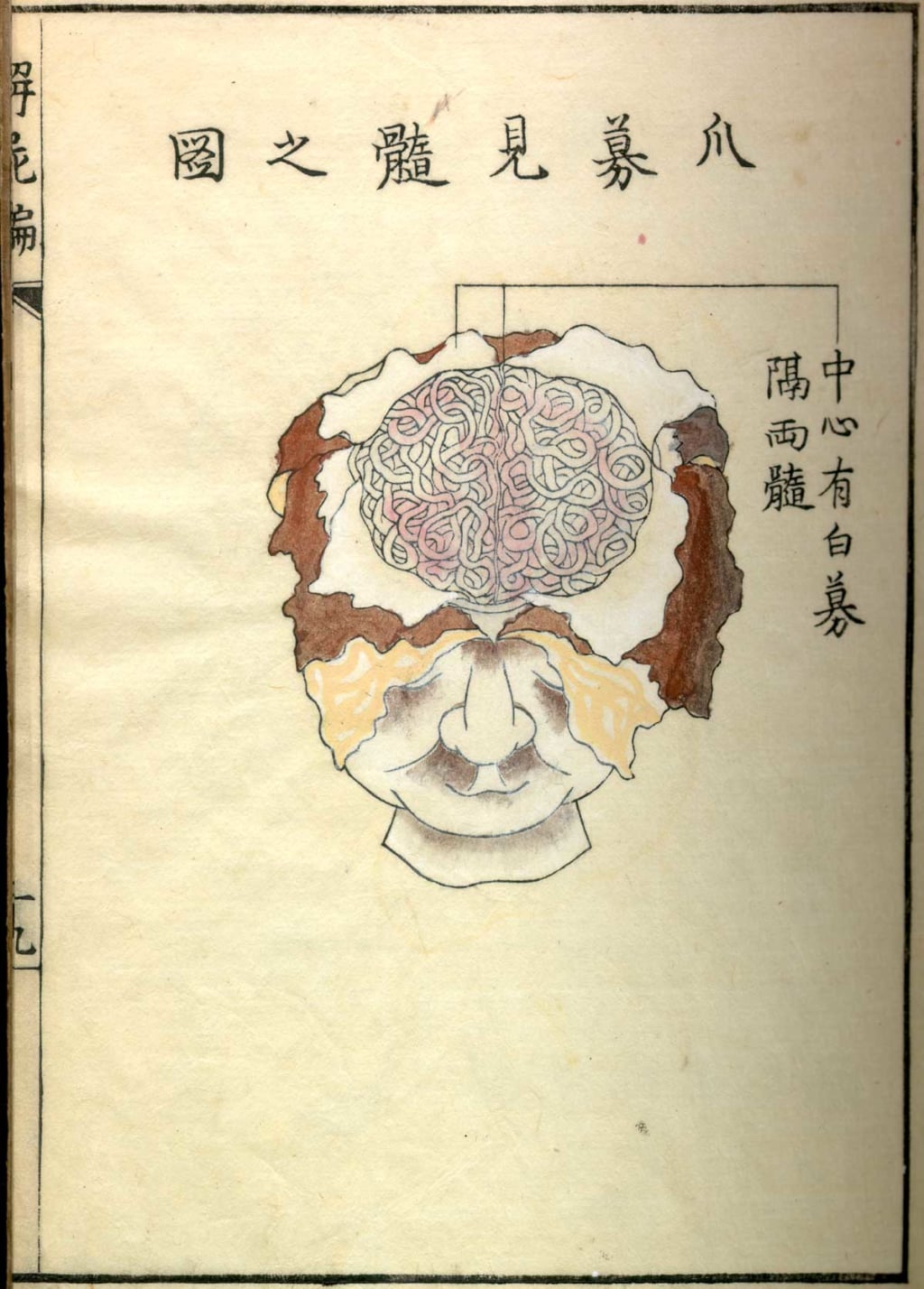
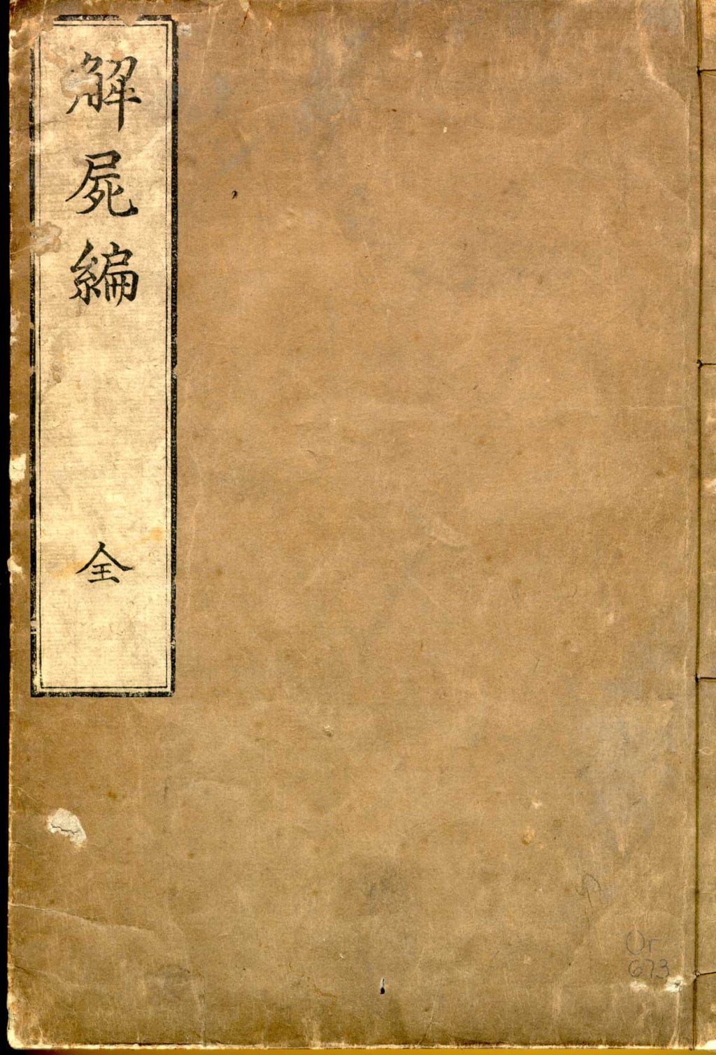
The illustrations engraved on wood in 'Kaishi Hen' are the vestiges of the first human dissections that took place in Japan.
© Public Domain
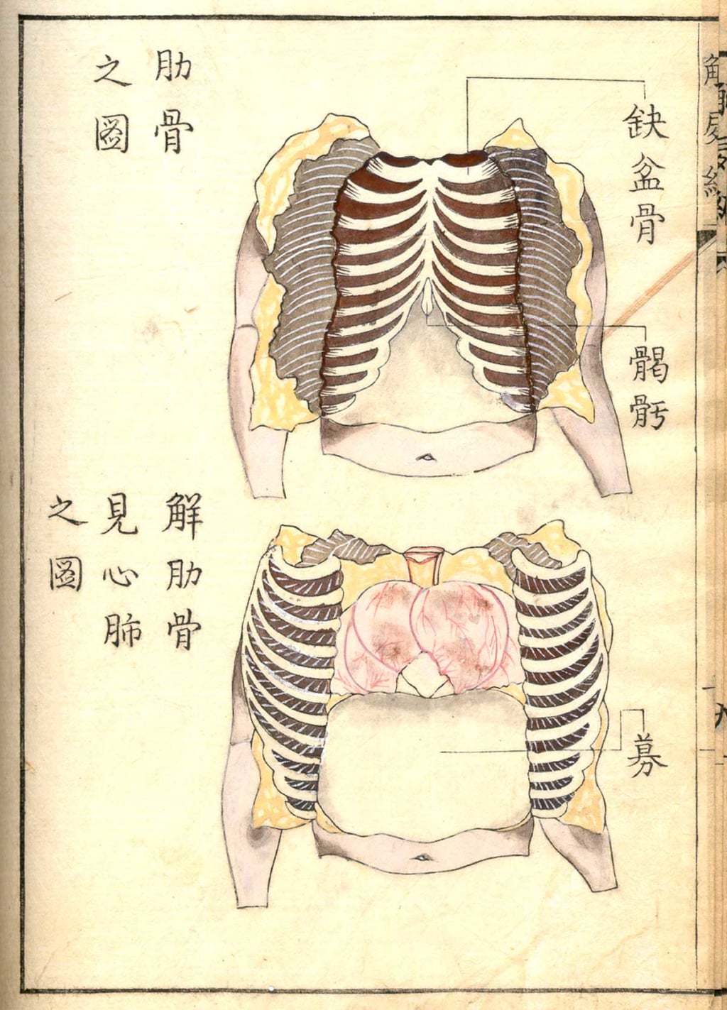
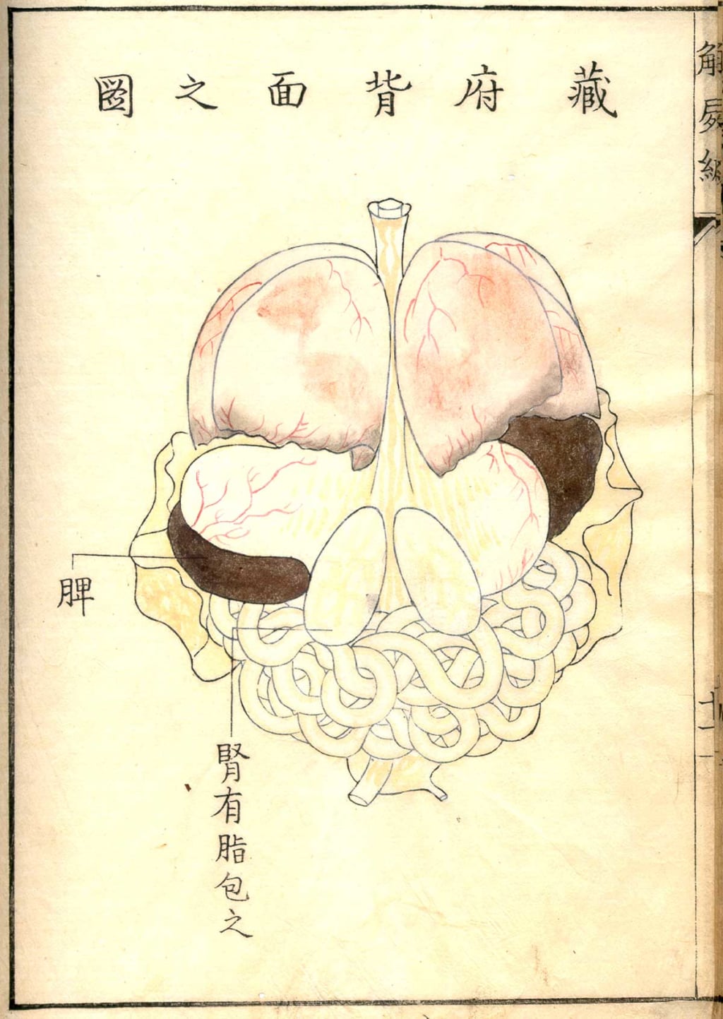
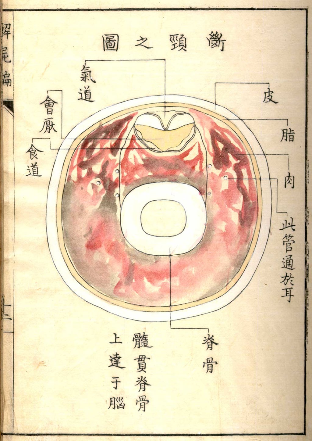
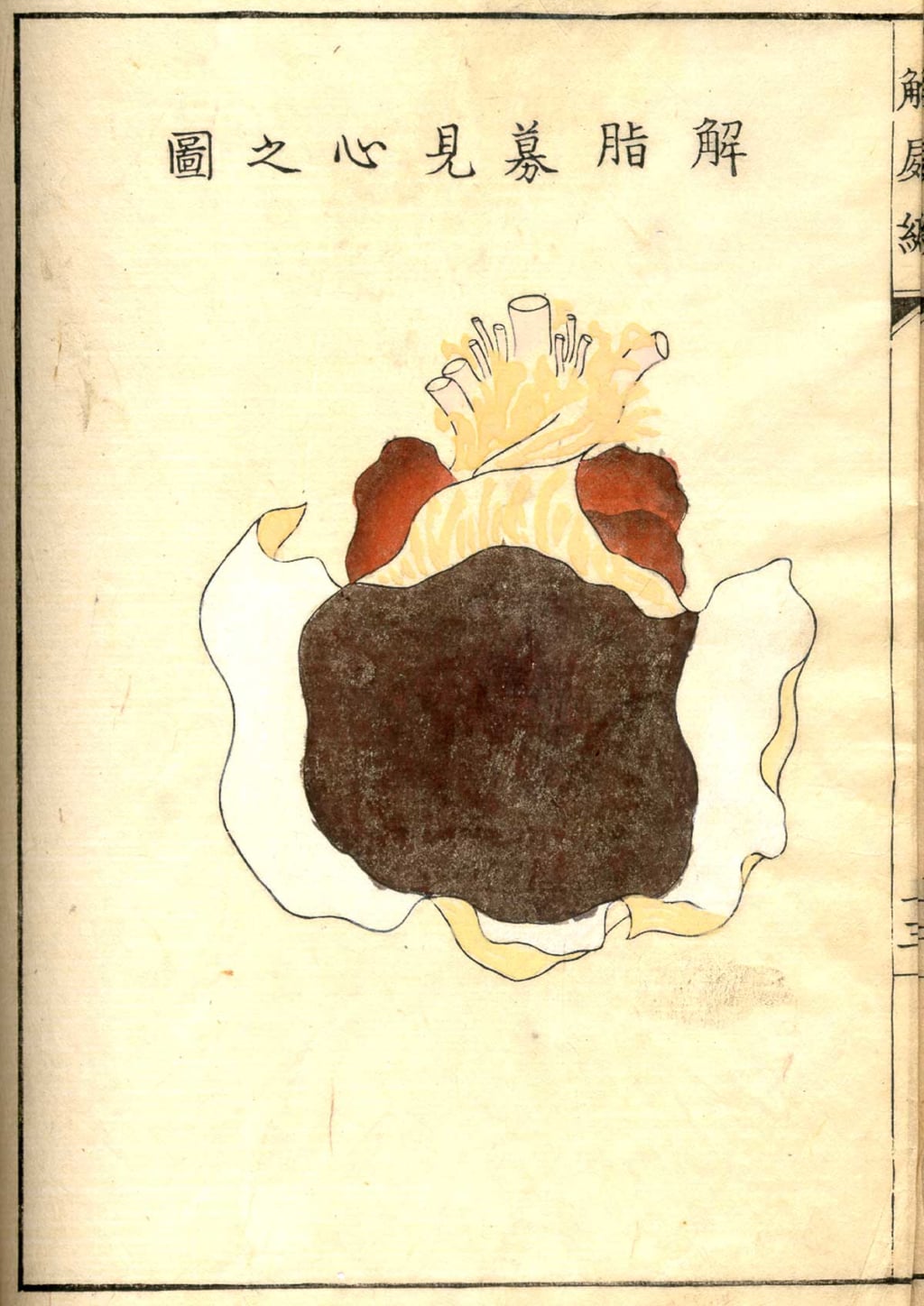
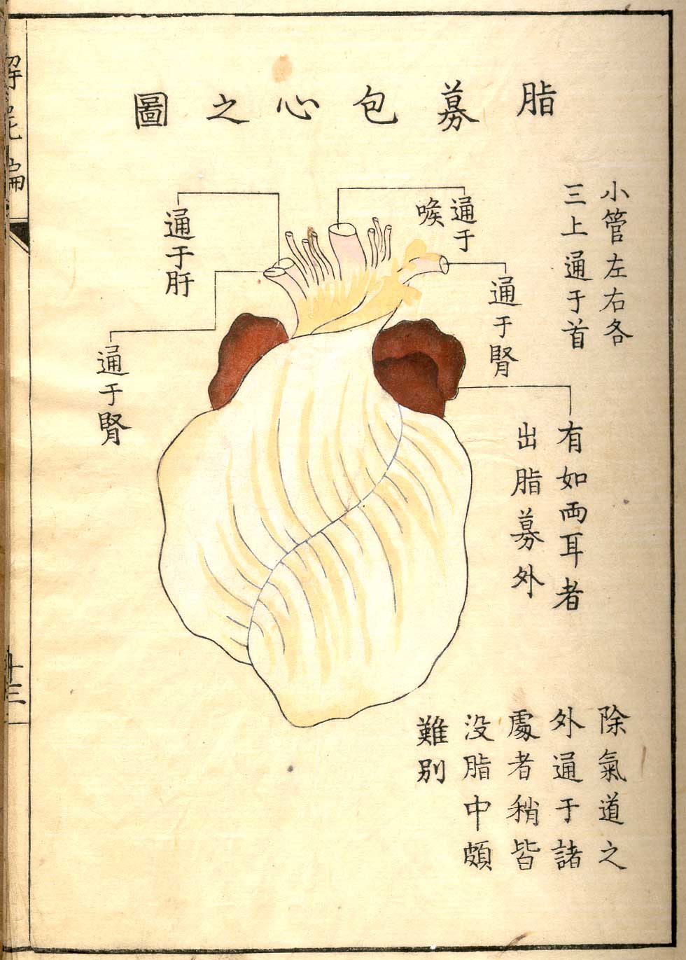
This series of 23 illustrations, compiled in an anatomical atlas entitled Kaishi Hen, represents various cross-sections of the human body. These hand-coloured wood engravings are the work of Japanese painter and sculptor Aoki Shukuya, and are based on sketches by Shinnin Kawaguchi, a disciple of Gengai Ogino, a Japanese doctor who studied Western medical theory.
The beginnings of experimental medicine
This anatomical atlas, published in 1772, marked the beginnings of experimental medicine in Japan. Its illustrations of intestines, the brain, the skeleton, and lungs depict the dissection of the body of a criminal executed in Kyoto two years earlier. At the time, this was only the third human dissection to have been carried out in Japan, the first having taken place in 1750 and also having been documented through wood engravings, carried out by artist Asanuma Suketsune and included in the book A Galaxy of Old Japanese Medical Books I. Dissection had been forbidden in the country since antiquity, and was only officially authorised in 1858.
Kaishi Hen (1772) is an anatomical atlas engraved on wood by Aoki Shukuya and digitised by the United States National Library of Medicine.
© Public Domain
© Public Domain
© Public Domain
© Public Domain
© Public Domain
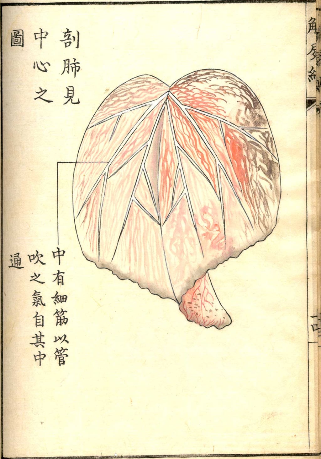
© Public Domain
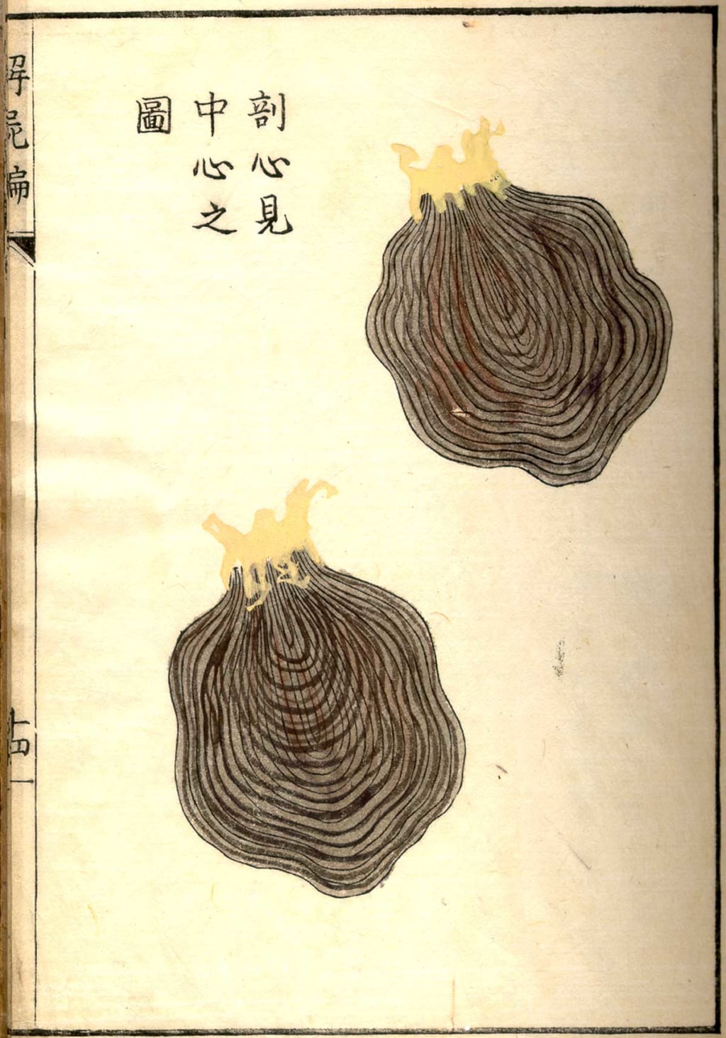
© Public Domain
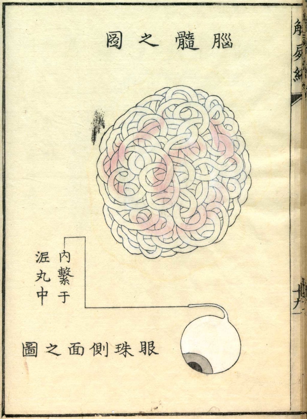
© Public Domain
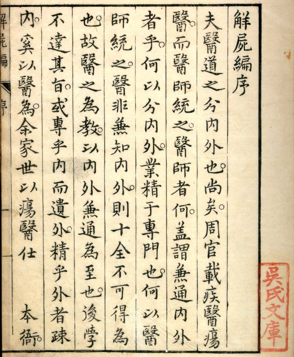
© Public Domain
© Public Domain
TRENDING
-
A House from the Taisho Era Reveals Its Secrets
While visiting an abandoned building, Hamish Campbell discovered photographs the owner had taken of the place in the 1920s.

-
The Taboo-Breaking Erotica of Toshio Saeki
The master of the 1970s Japanese avant-garde reimagined his most iconic artworks for a limited box set with silkscreen artist Fumie Taniyama.

-
With Meisa Fujishiro, Tokyo's Nudes Stand Tall
In the series 'Sketches of Tokyo', the photographer revisits the genre by bringing it face to face with the capital's architecture.

-
Masahisa Fukase's Family Portraits
In his series ‘Family’, the photographer compiles surprising photos in which he questions death, the inescapable.

-
Hajime Sorayama's Futuristic Eroticism
The illustrator is the pioneer for a form of hyperrealism that combines sensuality and technology and depicts sexualised robots.


